


‘Green Dentistry’ refers to the delivery of oral health care and dental treatments using technologies, procedures, and materials that promote environmental and planetary health. It incorporate high-tech innovations that enhance efficiency and effectiveness while reducing the amount of waste and pollution in the environment.
Klinik Pergigian Fauziah is always mindful of incorporating technologies, procedures, and practices to make the clinics eco-friendly. This means every facet of the practice of dentistry in our clinics are continuously upgraded as technology becomes available to incorporate a better, more efficient, and eco-friendly dental clinic.

One of the most significant innovations for Green Dental practices, is the digital radiography which eliminates the need for film X-rays. With digital X-rays, dental practices dramatically reduce harmful chemicals such as lead and silver that are released into the environment. Patients are exposed to approximately 90% less radiation with digital imaging compared to conventional X-rays. Also, images are available to the dentist immediately, are of better quality and can be enlarged for greater diagnostic detail and accuracy.
We use high-quality, biodegradable disinfectants, and steam sterilisation methods that doesn’t require ventilation for chemical vapours, or a hazardous waste permit or the disposal of toxic chemicals into our water supply. We increase as much possible the use of cloth bibs, gowns, and supplies that are washable and are able to be disinfected, so as to be reused. This eliminates the use of disposable paper products such as paper cups.
In these current times most people are interested in natural looking restorations. Eco-dentistry is in line with adhesive, metal-free dental restorations using direct porcelain veneers or porcelain veneers which only need minimal or no preparation. Another option are full ceramic crowns. They provide metal-free, aesthetic smile treatments, help eliminate heavy metal waste associated with silver amalgam fillings that could end up in water supplies.
Because patients often prefer adhesion dentistry, tooth-coloured restoration benefits the environment as well as the health of the patient by getting rid of heavy metal waste from main water supplies. To this end, Klinik Pergigian Fauziah has not used silver amalgam restorations since 2006.
Experience Precision, Comfort, and Speed in Your Dental Visits
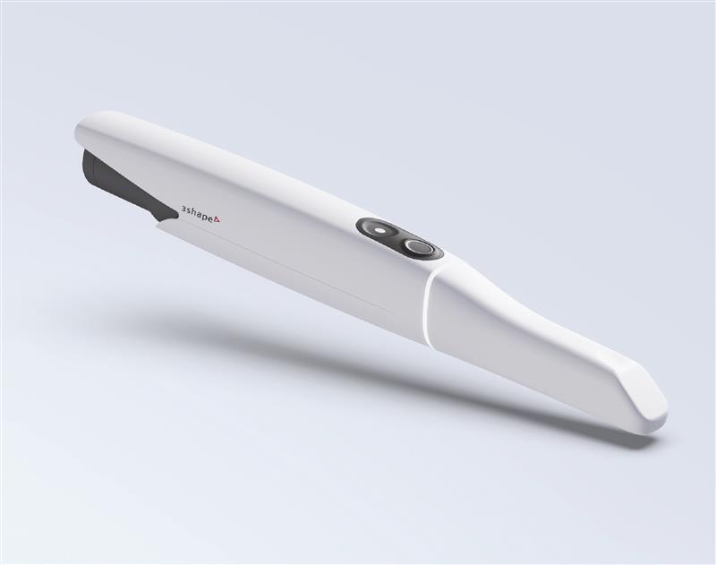
Say goodbye to messy impressions and hello to modern dentistry with our Intraoral Digital Scanner – the latest in dental scanning technology now available at our clinic.
What Is an Intraoral Digital Scanner?
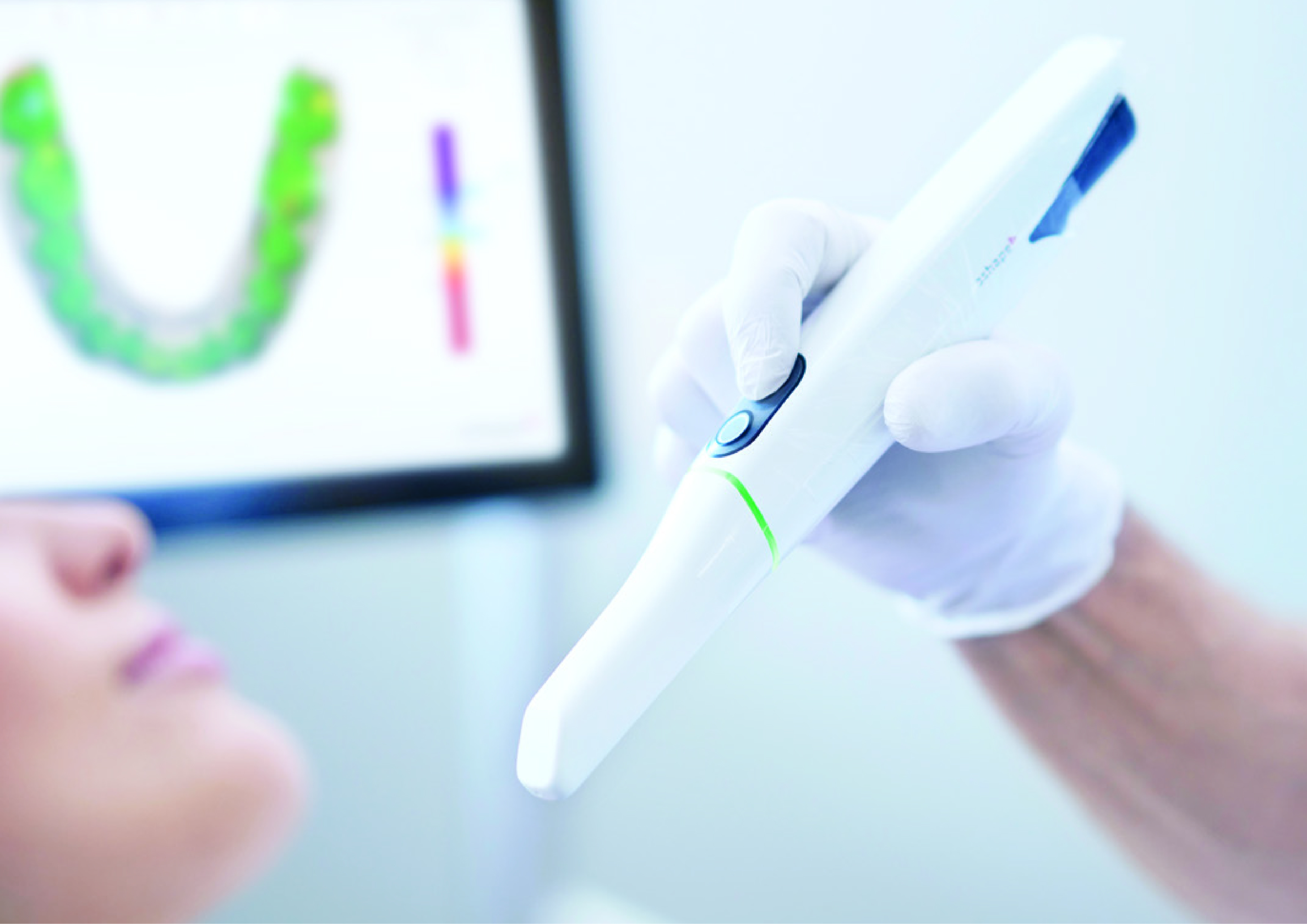
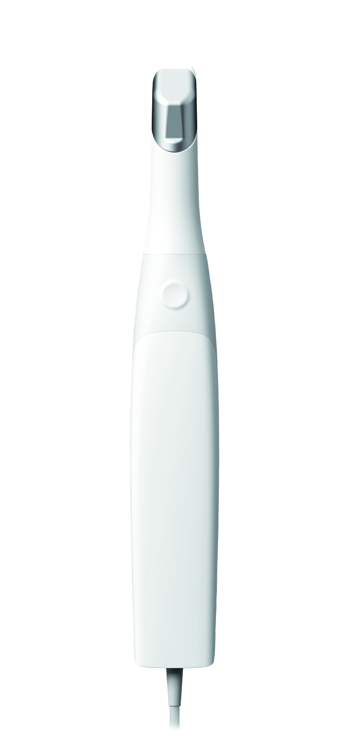
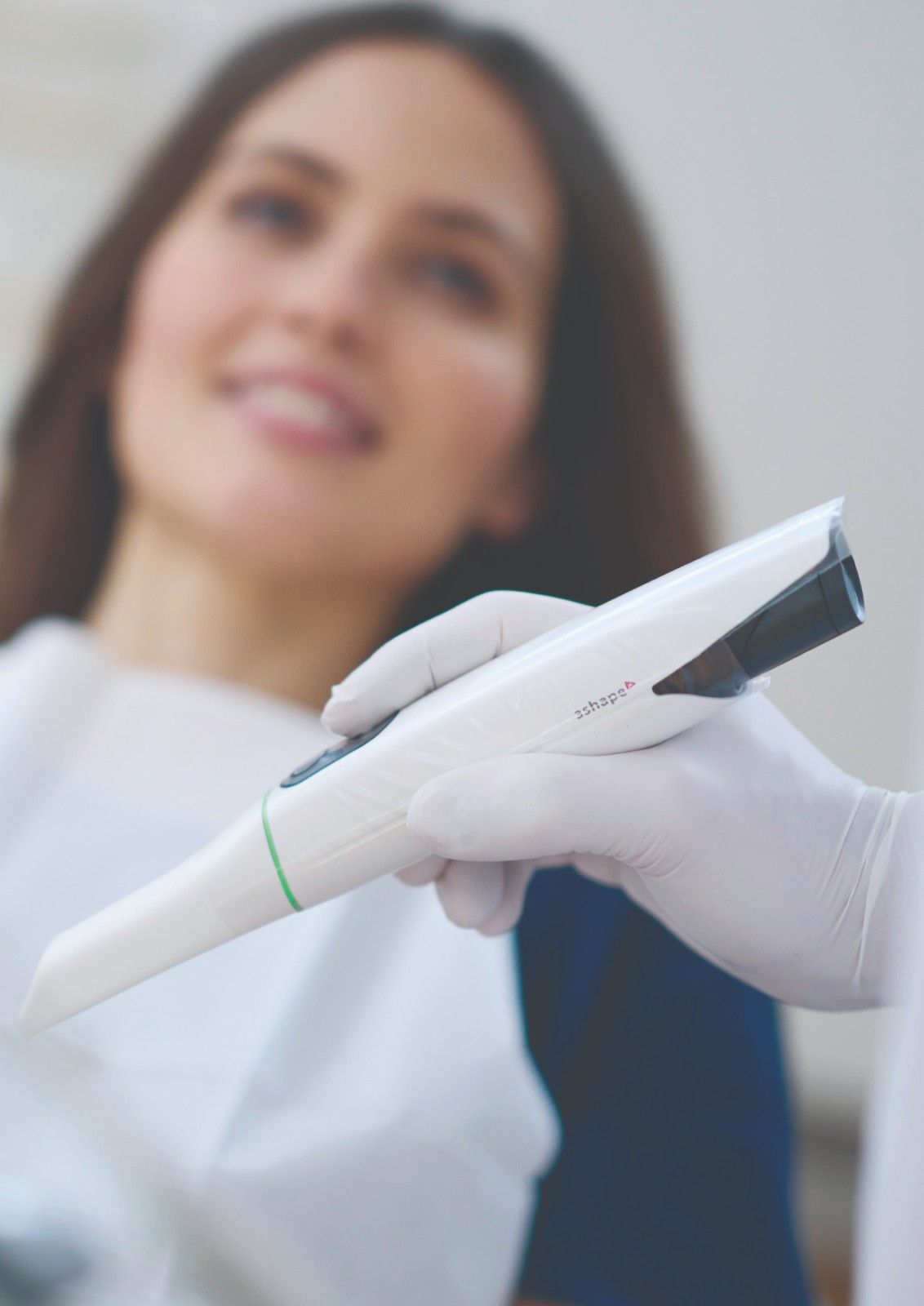
An Intraoral Digital Scanner is a handheld device that captures high-resolution 3D images of your teeth, gums, and bite in real time. It replaces traditional impression materials with fast, accurate digital imaging – giving you a more comfortable and efficient experience.
• The scanner uses a small, wand-like camera to take thousands of pictures per second.
• These images are stitched together into a precise 3D model of your mouth.
• Your dentist can review the scan instantly and share the results with you on-screen.
No Messy Impressions
Forget about biting into trays filled with goo. The digital scan is clean, quick, and comfortable.
Faster Appointments
Digital impressions are captured in minutes, and results are available instantly.
Exceptional Accuracy
High-definition 3D scans reduce errors and improve the fit of crowns, aligners, and other dental appliances.
Better Communication
You can see what your dentist sees, helping you better understand your treatment plan.
Eco-Friendly
Digital scans reduce the need for disposable impression materials, making this a more sustainable choice.
.jpg)
Our Intraoral Scanner is commonly used for:
• Digital impressions for crowns, bridges, and veneers
• Invisalign®️, Angel Aligner and other clear aligner treatments.
• Implant planning and restorations
• Monitoring changes in your bite or tooth wear
• Baseline records for ongoing dental care
DIGITAL DENTAL RADIOGRAPHY
In Our Efforts To Increasingly Go “Green” In Delivering Oral Health Care And Dental Treatment, We Chose To Use Digital Dental Radiographs (Digital X-Rays) To Better Detect, Diagnose, Treat, And Monitor Oral Conditions And Diseases.
Digital radiography is a type of X-ray imaging that uses digital X-ray sensors to replace traditional photographic X-ray film, producing enhanced computer images of teeth, gums, and other oral structures and conditions. This means the need for traditional film X-ray systems is no longer required. With digital X-rays, we dramatically reduce the harmful chemicals such as lead and silver that are released into the environment.
Additionally, patients are exposed to less radiation with digital imaging compared to traditional X-rays. Also, images are available to us immediately, are of better quality than traditional X-rays and can be enlarged for greater diagnostic detail and accuracy.
Digital dental images are acquired through 3 methods: the direct method, indirect method and semi-indirect method. The direct method uses an electronic sensor placed in the mouth to record images. The indirect technique uses an X-ray film scanner to view traditional dental X-rays as digital images. The semi-indirect digital technique combines a sensor and scanner to convert dental X-rays into digital film.

Digital dental radiographs can be taken inside (intraoral) or outside (extraoral) of the mouth. Intraoral X-rays, the most commonly taken dental X-ray, provides great detail and are used to detect cavities, check the status of developing teeth, and monitor teeth and bone health.
Extraoral X-rays do not provide the detail of intraoral X-rays, and are not used to identify individual tooth problems. However, they are used to detect impacted teeth, monitor jaw growth and development, and identify potential problems between teeth, jaws and temporomandibular joints, or other facial bones.
TYPES OF INTRAORAL X-RAYS INCLUDE:
• Bitewing X-rays, which are taken with the patient biting down on film, showing details of the upper and lower teeth in one area of the mouth. Each bitewing shows a tooth from its crown (top) to the level of the supporting bone. Bitewing X-rays are used to detect decay between teeth and changes in bone density caused by gum disease, as well as to determine the fit of dental crowns or restorations, and the marginal integrity of tooth fillings.
• Periapical (limited) X-rays show the whole tooth from the crown to beyond the root tips, to the supporting bone in one area of either the upper or lower jaw. Periapical X-rays are used to detect root structure and surrounding bone structure abnormalities. Showing bone loss around each tooth, periapical X-rays aid in treating conditions such as periodontitis, advanced gum disease, and detecting endodontic lesions (abscess).
TYPES OF EXTRAORAL X-RAYS INCLUDE:
• Panoramic (panorex) X-rays, which require a machine that rotates around the head, show the entire mouth including all the teeth in the upper and lower arch in one image. Panoramic X-rays are used to plan treatment for dental implants, detect impacted wisdom teeth and jaw problems, and diagnose bony tumours and cysts.
• Cephalometric projections, which show the entire head, help examine teeth in relation to a patient's jaw and profile. Orthodontists, specialists in aligning and straightening teeth, use cephalometric projections to develop their treatment plans.
• Cone beam computerised tomography (CBCT) shows the body's interior structures as a three-dimensional image. CBCT, often performed in a hospital, is used to identify facial bone problems, such as tumours or fractures.
• CT scans are also used to evaluate bone for dental implant placement and difficult tooth extractions, to avoid possible complications during and after surgical procedures.
• The CBCT, which requires more radiation than panoramic radiographs, does not slice images. Instead, its cone-shaped beam scans on both the upper and lower mouth areas at once. The data is captured by a 2-Dimensional array and creates high-resolution images, then combined to form a 3-D image of the bony structures.
Klinik Pergigian Fauziah has 2 of these machines, the Sirona Orthopos which we use in treatment planning for all surgical procedures like minor oral surgery to remove impacted wisdom teeth, and in detailed planning for placements of dental implants.
Benefits of digital dental radiographs compared to traditional dental X-rays include the following:
• Digital radiographs reveal small hidden areas of decay between teeth or below existing restorations (fillings), bone infections, gum (periodontal) disease, abscesses or cysts, developmental abnormalities, and tumours that cannot be detected with only a visual dental examination.
• Digital radiographs can be viewed instantly on any computer screen, manipulated to enhance contrast and detail, and transmitted electronically to specialists without quality loss.
• Early detection and treatment of dental problems can save time, money, and discomfort.
• Digital micro-storage technology allows greater data storage capacity on small, space-saving drives.
• Dental digital radiographs eliminate chemical processing and disposal of hazardous wastes and lead foil, thereby presenting a "greener" and eco-friendly alternative.
• Digital radiographs can be transferred easily to other dentists with compatible computer technology, or photo printed for dentists without compatible technology.
• Digital sensors and PSP (photostimulable phosphor) plates are more sensitive to X-radiation and require 50 to 80 percent less radiation than film. This technology adheres to the ALARA (As Low As Reasonably Achievable) principle, which promotes radiation safety.
• Digital radiograph features, including contrasting, colorising, 3-D, sharpness, flip, zoom, assist in detection and interpretation, which in turn assist in diagnosis and patient education. Digital images of problem areas can be transferred and enhanced on a computer screen next to the patient's chair.
• Digital dental images can be stored easily in electronic patient records and sent quickly electronically to insurance companies, referring dentists or consultants, often eliminating or reducing treatment disruption leading to faster dental insurance reimbursements.
Most digital sensors and PSP plates can't be sterilised and therefore require protective plastic barriers that must be changed between patients to prevent cross contamination and infection.
While radiation exposure is low with digital radiographs, no one should receive more radiation than absolutely necessary. Protective lead aprons and thyroid collars are routinely used especially for pregnant women, women of childbearing years, and children.
It is safe for pregnant women to have up to 4 radiographs per office visit, although most patients and doctors will elect to delay radiographs until the pregnancy is over. There should be no concerns for a pregnant woman to have an X-ray taken in an emergency situation. Precautions such as the use of ‘double lead aprons’ cut radiographic exposure down to nearly immeasurable levels. Women who are breast-feeding or trying to become pregnant do not need to delay X-rays.

